
Articles
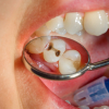
In this work we demonstrate the suitability of confocal Raman microscopy for the characterisation of carious dental tissues. Samples of enamel and dentine, presenting carious lesions in different stages of progression, were evaluated by comparing the depolarisation ratio of the PO43– symmetric stretching band at 959 cm–1 in the different tissues. Both line and area scans were performed to gauge these variations. Moreover, the obtained results were compared with the tissues’ behaviour when interacting with ultraviolet radiation, namely the induced fluorescence in some tissues. The depolarisation ratio has proven to be a valuable tool in recognition of demineralisation of both enamel and dentine due to caries. The analyses of the collagen bands in the dentine sample turned out to be more difficult to evaluate due to high fluorescence in the carious region.
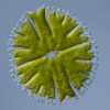
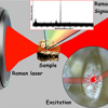
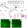
Different spectroscopic techniques have been combined to provide additional and complementary information for decades. Increasingly, this is being expanded beyond just two techniques and may include spatial/imaging information as well. All of which bring their own challenges. In “Multimodal imaging of cells and tissues: all photons are welcome”, David Perez-Guaita, Kamila Kochana Anja Rüther, Phillip Heraud, Guillermo Quintas and Bayden Wood report an example of these new approaches. They look at the use of infrared, Raman and X-ray fluorescence spectroscopies to obtain combined imaging data of whole algal cells and discuss how to overcome the challenges.
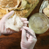
Edible bird’s nest (EBN) is a highly valued food, especially in China. Due to this, there is the potential to bulk out the EBN with artificial additives to receive a higher price. This article shows yet another way in which spectroscopy is used to detect adulteration of food and prevent fraud in a quick and cost-effective manner.
There are a number of advantages of Raman microspectroscopy, including its the ease of its application, as in many cases no or very little sample preparation is needed and the experiments are performed at atmospheric pressure.
It is not every issue that one of our articles starts with a quotation in medieval English, and it is appropriate as two of our articles cover the use of spectroscopy in cultural heritage. This is yet another field where the rich information provided by spectroscopy, along with its non-destructive nature (for many techniques), portability and ability to generate chemical images make it the answer to many questions. Kate Nicholson, Andrew Beeby and Richard Gameson are responsible for the medieval English at the start of their article “Shedding light on medieval manuscripts”. They describe the general use of Raman spectroscopy for the analysis of historical artefacts, and, in particular, their work on medieval European manuscripts and 18th century watercolour pigments. They stess the importance of checking the actual laser power density to avoid damage to priceless artefacts.
This article looks at the use of Raman and XRF spectroscopies to investigate the different deterioration processes caused by marine aerosols. These techniques can detect the decay compounds and the original composition of the different materials from historical buildings close to the sea, which can then be used to explain the reactions that take place on them. This helps in the development of remedial actions and preventive conservation strategies for historical buildings.

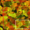
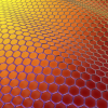
As you will have noticed from this issue’s cover, we are making a colourful start to 2016. In the first article on “The analytical niche for Raman spectroscopy in biological pigment research”, Daniel Thomas and Cushla McGoverin suggest that Raman spectroscopy may have a particularly valuable role in pigment biology research. Pigments are almost universal in biology and are the basis of much of what we find attractive in flowers, birds and sea life, such as the fan corals on the cover. The authors show how Raman spectroscopy can be used to quickly confirm the presence of a pigment as well as providing more detailed knowledge about unknown pigments.
Graphene has been receiving a large amount of interest as its commercial possibilities begin to be realised. Now, with hundreds of companies offering commercial graphene production, analytical measures of graphene quality are required. Raman spectroscopy can be used to “understand the number of layers, strain, doping and importantly the level of disorder present in graphene”, which is described in this article: “Graphene characterisation and standardisation via Raman spectroscopy” by Andrew Pollard and Debdulal Roy.
In the Tony Davies Column, we learn about “Automated detection of counterfeit drugs using multimodal spectroscopy and advanced web-based software platforms”. With the increase in trafficking of counterfeit medicines and other products, there is a need for definitive results from an on-site analyser useable by customs officers. The German authorities have commissioned the development of a multi-modal, transportable inspection system, including intelligent data processing and evaluation, for fast spectroscopic recognition of illicit drugs and counterfeit medicines. This is described in the column.
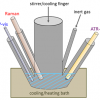
In situ spectroscopic methods such as infrared, Raman and UV/vis spectroscopy are powerful tools to gain insight into reaction mechanisms and catalyst actions in homogeneously catalysed reactions. These methods and combinations of them offer great potential for the real-time monitoring of reactions in the liquid phase, for mechanistic studies as well as process control and kinetics.
“In vivo Raman spectroscopy of skin” is Paul Pudney’s topic. The skin is a most important part of our bodies. There is great interest in studying it to help understand the many skin diseases we are prone to, including cancer, to develop skin care products and, increasingly, as an alternative route to administer pharmaceuticals instead of through the gut. Raman spectroscopy is an excellent tool to study these, and has particular advantages in its ability to do so in vivo.
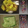
The “Application of Raman and photoluminescence spectroscopy for identification of uranium minerals in the environment” is described by Eric Faulques, Florian Massuyeau, Nataliya Kalashnyk and Dale Perry. Uranium forms a large number of compounds and complexes, and these are most helpful in the study of uranium, its chemistry and transport in the environment. Raman and photoluminescence spectroscopy provide complementary information and are powerful tools for direct speciation of uranium and identification of natural uranyl minerals relevant to the environment.
“Optical spectroscopy in therapy response monitoring: an awakening giant” by Arja Kullaa, Surya Singh, Jopi Mikkonen and Arto Koistinen looks at the important advances made by optical spectroscopy techniques, such as diffuse optical spectroscopic imaging (DOSI), Raman, diffuse reflectance and fluorescence spectroscopy, in changing how cancer is managed in patients. The ability to repeatedly monitor tumour dynamics to see how effective a particular treatment has been has enormous potential for us all.
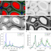
“Elucidating structural and compositional changes in plant tissues and single cells by Raman spectroscopic imaging” is the topic of the next article by Batirtze Prats Mateu, Barbara Stefke, Marie-Theres Hauser and Notburga Gierlinger. Understanding plant cells is important for the best use of plants in traditional and new applications. Raman spectroscopic imaging represents one of the best ways to unravel the molecular structure in the native environment of plant tissues.
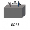
“Emerging sampling approaches for Raman analysis of foods” by Nils Kristian Afseth, Matthew Bloomfield, Jens Petter Wold and Pavel Matousek describes how a number of instrumental developments are enabling Raman spectroscopy to find increasing applications in food analysis. They show how Spatially Offset Raman Spectroscopy (SORS) is being used to analyse quality parameters in salmon, including the content of fat, its fat composition and the content of carotenoids. Traditionally, the preserve of NIR spectroscopy, Raman may increasingly be used for the analysis of food and other biological matrices.
Malvina G. Orkoulaa and Christos G. Kontoyannisa,b
aDepartment of Pharmacy, University of Patras, Rio-Patras, Greece. E-mail: [email protected]
bICE-HT/FORTH, PO Box 1414, University Campus, Rio-Patras, Greece
The single cell Raman spectrum (SCRS) enables cell probing and sorting to study phenotypes and ecophysiology of single cells and explore individual cells in situ in a label-free and non-destructive manner.
In resonance Raman scattering (RRS), the amount of structural and chemical information deduced can be increased by analysing the polarisation of the inelastically scattered light, including the degree of molecular aggregation in bio-molecules in their natural environment.
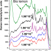
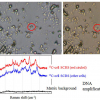
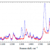
Thiabendazole (TBZ) is a chemical fungicide and parasiticide used to prevent mould, blight and other diseases resulting from long transportation and storage, largely used as an ingredient in waxes applied to the skins of citrus fruits. The authors describe their work using near infrared-surface-enhanced Raman spectroscopy and conventional Ag nanoparticles, which showed that TBZ was found both on organic fruit and at levels higher than regulations allow.
Tip-enhanced Raman spectroscopy (TERS) and tip-enhanced Raman mapping (TERM) can be used advantageously to investigate the carbon allotropes graphene and single-walled carbon nanotubes, with a spatial resolution in the nanometre range
