Patrick Garidel
Boehringer Ingelheim Pharma GmbH & Co. KG, Process Science, Protein Science, D-88397 Biberach an der Riss, Germany. E-mail: [email protected]
Introduction
During the last three decades a number of biologicals were developed and successfully commercialised due to their high therapeutic effects and benefits. These achievements were mainly driven by the impressive advances in recombinant DNA and protein science technologies. Biologics are complex molecular entities manufactured via biotechnology processes using mammalian or bacterial cell culture technologies.1,2 The most common class of biologics are monoclonal antibodies, whereas other therapeutic biologics belong to the interferon family, or are insulin or insulin-like entities with derivatives thereof or plasminogen activator proteins, to name just a few examples.
Compared to classical new chemical entities (NCEs), biologics are more sensitive with regard to stability, due to their large molecular weight and size, but also due to their complex three-dimensional structure that mainly determines biological activity.3,4 Very small changes in the three-dimensional structure may strongly influence biological activity. In addition to chemical instabilities, such as de-amidation, oxidation, isomerisation of certain amino acids, hydrolysis, disulfide scrambling or glycation, physical and colloidal instabilities such as protein denaturation or the formation of intrinsic and extrinsic protein particles are of high relevance due to the potential immunogenicity of protein aggregates.4,5 In addition to these challenges, the process itself can have a large impact on protein quality and integrity.
During the development of biologics, various clone candidates are screened and selected based on a number of quality attributes. Among theses, cell viability, cell density, genetic stability or cell productivity are important parameters. Furthermore, the expressed therapeutic protein has to be analytically characterised as much as possible in addition to determining its stability. A main challenge is the extremely small amount of protein available during the early research and development phase for the investigation of these selected parameters.
To investigate and screen the stability of proteins in solution, various biophysical methods are used, which rely on monitoring stability indicating parameters. The most common methods are calorimetry, fluorescence spectroscopy (intrinisic or extrinsic), infrared spectroscopy or circular dichroism.6,7,8
The stress applied to the protein solutions is, in most cases, a specific temperature scan, thus monitoring conformational and thermal stability, which to a first approximation is correlated to overall long term stability. One has to mention that, during such thermal-induced, fast-track stability screening experiments, chemical degradations, such as de-amidation or oxidation are not directly monitored or considered. Thus, only a specific view of stability is considered, namely thermostability.9
Depending on the techniques used, the registered stability indicating parameter is quite different, thus monitoring different aspects of protein thermostability and temperature-induced unfolding. For example, during a calorimetric experiment, the heat capacity curve is registered, which shows various enthalpic contributions during protein unfolding, whereas using infrared spectroscopy changes in the secondary structure are monitored. For more details see references.6,10 However, not each of these methods can be applied in the early research and development phase due to technical restrictions as well as a lack of available material for stability screening. Among the above cited methods, fluorescence spectroscopy is certainly the method of choice if just very small amounts of materials are available. Usually, the fluorescence experiments are performed as intrinsic or extrinsic fluorescence thermostability screening. Using external fluorescence dyes, which are often hydrophobic, one has to be aware of possible dye–protein interactions that may lead to partial protein denaturation. In cases for which this is an issue, intrinsic fluorescence spectroscopy is more appropriate and reliable, especially for proteins containing tryptophan, tyrosine or phenylalanine (see below).
Nevertheless, using the same fluorometric equipment, it is also possible to perform another thermostability indicating assay, namely right angle light scattering (RALS), which is a label-free method.
This article describes the method and presents examples of how this assay can be used during early research and development phase in protein science.
Experimental
Material
The used monoclonal antibodies (mAb) were produced by mammalian cell culture technology and the single chain antibody (SCA) fragment by Escherichia coli cell culture technology.1 The proteins were recovered, purified and characterised according to procedures described in the literature.1,2,6,11 Protein monomer content was >99% (based on high performance size exclusion chromatography measurement).
Right angle light scattering (RALS)
RALS stability screening was performed on a standard fluorescence spectrometer (for example, PTI Luminescence QuantaMaster Spectrometer, Photon Technology International Ford, UK) equipped with a self-manufactured, peltier-controlled, multi-sample holder. Excitation is performed at λExc = 320 nm and emission recorded at λEm = 320 nm.
Intrinsic tryptophan fluorescence (ITF)
ITF experiments were performed with the same equipment as described previously.12
Results
In the used set-up, intrinsic fluorescence (IF) and RALS are monitored concurrently during the same experiment. For ITF (intrinsic tryptophan fluorescence) λExc = 295 nm and emission is recorded between λEm = 300 nm and 400 nm as described recently.12 However, RALS determination uses an excitation and emission wavelength of 320 nm, which is recorded by the detector at 90°. During the stability screening experiment, the protein solution is temperature ramped from 20°C to 90°C using a specific temperature protocol.
IF measures possible environmental and/or structural changes for specific amino acids such as tryptophan, tyrosine and phenylalanine (depending on λExc).13 The use of tryptophan as a fluorophore is based on the fact that its emission is highly dependent on the polarity of the fluorophore environment. Thus for ITF, potential changes in tryptophan environment arising from stress-induced conformational alterations are monitored and plotted as a function of temperature as intensity ratios such as 350/330 (for more details see cited References 12 and 13).
The RALS assay is employed to detect and monitor subtle alterations in associative behaviour of stressed proteins in otherwise visually clear solutions. RALS is able to investigate macroscopic changes in a usually soluble protein molecule transition to the formation of insoluble protein particles.
Figure 1 shows examples of RALS measurements monitored as a function of temperature. In general, one observes, at a specific temperature a rapid increase in intensity for the RALS signal. This is related to thermal-induced instability of the protein in solution due to associative properties of the protein, followed by the potential formation of larger particles, which are formed upon the unfolding of the protein. The RALS data of six experiments of two samples are presented in Figure 1. For sample A (A)–(C), a relatively sudden transition at 61°C is observed. This temperature is set as the aggregation temperature (Tagg). Different approaches can be used for the definition of this temperature, for example by the determination of the temperature at which the curve suddenly increases and leaves the baseline (onset), or the inflection point of the S-shaped curve can be determined and used. Independent of which method is applied, for comparative studies, the same evaluation procedure has to be used. In the presented examples, both data evaluation methods lead to very similar results (see Figure 1).
 temperature profile showing inter- and intra-sample reproducibility. Samples 1A–1C show inter-sample reproducibility, i.e. three independent samples, for a monoclonal antibody (mAb) at pH 5.0 in a saline-acetate buffer, whereas samples AA–AC are examples for intra-sample reproducibility, i.e. same sample, but three independent measurements for mAb at pH 7.0 in a saline-phosphate buffer.")
The observed transition in the RALS experiment is indicative of the formation of some type of association phenomenon, induced by temperature stress. In certain cases, at the end of the temperature profile, the protein sample may form white flocculent protein precipitates. An example is shown in Figure 1 for inter-sample reproducibility, i.e. the investigation of three independent samples for the same solution conditions (samples 1A, 1B and 1C, Tagg = 74°C +/– 0.2°C) as well as an example of the investigation of the same sample (intra-sample), however, three independent RALS experiments (samples AA, AB and AC, Tagg = 61°C +/– 0.3°C) (remark: the pH of the solutions of sample 1 and sample A are different). These two examples show a high reproducibility of the thermal-induced stability screening assay.
Fluorescence-based methods have some advantages over other methods such as calorimetry for thermal-induced stability screening.14 For calorimetric experiments, usually ca 1 mg of protein is required for one experiment. With the same amount of protein, depending on the used set-up, about 100 fluorescence experiments can be performed, i.e. about 100 different solution conditions can be investigated. Furthermore, due to the high sensitivity of fluorescence-based methods and the extremely low amount of material required, these methods are greatly suited to thermostability evaluation during the early research development phase.
For example, during the early development phase various clones are assessed and ranked. Clones are evaluated with regard to various parameters, for example, the productivity of the clones, as well as protein characteristics of the expressed protein, for example, binding capacity. However, other protein parameters such as glyco profile (for glyco proteins), solubility, thermodynamic stability or aggregation propensity have also to be evaluated. For instance, Figure 2 shows a series of monoclonal antibody (mAb, ca 150 kD) candidates that have more or less the same binding capacity. Thus, based on this evaluation parameter alone, the different clones are very similar, however, the RALS assay clearly shows that their thermostability is quite different for the investigated clone candidates for the same solution conditions (Figure 2). The highest thermostability, i.e. highest Tagg, is detected for clone candidates 1, 5 and 7 with Tagg between 76°C and 78°C. Furthermore, one also observed that clone candidate 3 shows a higher turbidity in the presented solution compared to the other candidates.
. The measured binding activity for the seven proteins is very similar. For the RALS experiment, the isolated proteins were dialysed against the same buffer composed of 150 mM NaCl, 20 mM histidine buffer at pH 6.5 and investigated with regards to thermal induced aggregation.")
As well as thermostability screening of clone candidates, RALS is also suited for the evaluation of solution conditions during downstream and formulation development. For instance, Figure 3(a) shows the impact of ionic strength (sodium chloride) on Tagg of a single chain antibody (SCA, ca 30 kD) fragment. The basic formulation is composed of 15 mM acetate buffer at pH 5.5 and various amounts of sodium chloride starting from 0 mM up to 500 mM. A careful examination of the RALS curves show that, with increasing the amount of salt, Tagg increases. This shows that the presence of salt induces a conformational “stabilisation” of the protein structure and, as a consequence, Tagg increases from 54 (0 mM NaCl) to 63°C (75 mM NaCl) [Figure 3(b)]. However, with increasing salt concentration above 75 mM NaCl, Tagg decreases and at a high salt concentration of 500 mM NaCl, Tagg shows its minimum. Furthermore, at this high salt concentration high turbidity is also observed, indicating lower protein solubility and higher propensity for the formation of protein particles.
 RALS temperature profile for the evaluation of the thermal-induced aggregation of a single chain antibody (SCA) fragment as a function of ionic strength (0–500 mM NaCl) at pH 5.5 in 15 mM acetate buffer. (b) Tagg versus ionic strength plot for the investigated SCA fragment showing the impact of ionic strength on thermal aggregation stability.")
This assay can also be used during an iterative formulation development programme for the evaluation of excipient with regard to protein stability. The use of multi-sample set-ups permits a high number of samples to be tested in “parallel”. Currently developed instruments, using a RALS approach, allow the investigation of nearly 50 samples within 4 h. By variation of the heating rate, experimental time can be further optimised.
Conclusions
Within the label-free biophysical assay investigating the thermostability of proteins in solution, RALS is a valuable alternative method to calorimetry or vibrational spectroscopy. A benefit of running a RALS experiment lies in the fact that it can be run on a classic fluorimeter and, depending on the instrumental set up, even in parallel to intrinsic fluorescence spectroscopy. A major benefit of fluorescence-based methods, as well as RALS, is that per sample the required absolute protein amount is in the range of a few microgram.
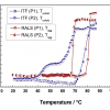
However, it is important to point out that the various available biophysical protein screening methods rely on monitoring different physical denaturation parameters (see discussion above). As a consequence, the observed denaturation temperature may be, for physical and biochemical reasons, different.14 Furthermore, depending on the used assay, different unfolding processes are observed and monitored. This is exemplified by a direct comparison of a RALS and ITF experiment for the same sample (Figure 4). During the ITF experiment, changes in the polarity environment of tryptophan are monitored. The ITF assay shows two transitions; one located at ca 50°C and a second at 70°C (Figure 4). Additional experiments have shown that the first transition is reversible (data not shown). However, in the RALS experiment, Tagg is observed at 73°C, monitoring a process of associative protein properties which, in most cases, are irreversible.

The comparative analysis of such data is very helpful to understand the stabilisation mechanism of the protein in solution as a function of, for example, added excipients, in order to develop a rational stabilisation strategy.
Acknowledgements
Inge Holl, Beate Fischer, Heidrun Schott and Michael Fäth are acknowledged for their excellent technical assistances and Joey Studts for critical reading of the manuscript.
References
- K. Bergemann, C. Eckermann, P. Garidel, S. Grammatikos, A. Jacobi, H. Kaufmann, R. Kempken and S. Pisch-Heberle, “Production and downstream processing”, in Handbook of Therapeutic Antibodies, Volume I: Technologies, Chapter 9, Ed by S. Dübel. Wiley-VCH, Weinheim, Germany, pp. 199–238 (2007).
- A. Jacobi, C. Eckermann and D. Ambrosius, “Developing an antibody purification process”, in Bioseparation and Bioprocessing, Ed by G. Subramanian. Wiley-VCH, Weinheim, Germany, pp. 431–457 (2007).
- P. Garidel and S. Bassarab, “Impact of formulation design on stability and quality”, in Quality for Biologics: Critical Quality Attributes, Process and Change Control, Production Variation, Characterisation, Impurities and Regulatory Concerns, Chapter 7, Ed by N. Lyscon. URCH Publishing, London, UK, pp. 94–113, (2008).
- M.C. Manning, D.K. Chou, B.M. Murphy, R.W. Payne and D.S. Katayama, “Stability of protein pharmaceuticals: an update”, Pharm. Res. 27(4), 544–575 (2010). doi: 10.1007/s11095-009-0045-6
- A.S. Rosenberg, “Effects of protein aggregates: an immunologic perspective”, AAPS J. 8(3), article 59, E501–E507 (2006).
- P. Garidel, W. Kliche, S. Pisch-Heberle and M. Thierolf, “Characterization of proteins and related analytical techniques”, in Protein Pharmaceuticals—Formulation, Analytics & Delivery, Ed by H.C. Mahler, G. Borchard and H. Lueßen. Chapter 2. Editio Cantor Verlag, Aulendorf, Germany, pp. 44–89 (2010).
- M. Weichel, S. Bassarab and P. Garidel, “Probing the thermal stability of MAbs by intrinsic tryptophan fluorescence. A practical approach for protein preformulation development”, BioProcess Int. 6(6), 42–52 (2008).
- P. Garidel and H. Schott, “Fourier-transform mid-infrared spectroscopy for the analysis and screening of liquid protein formulations. Part 2: Detailed analysis and applications”, BioProcess Int. 4(6), 48–55 (2006).
- H.J. Hinz, C. Steif, T. Vogl, R. Meyer, M. Renner and R. Ledermüller, “Fundamentals of protein stability”, Pure Appl. Chem. 65(5), 947–952 (1993). doi: 10.1351/pac199365050947
- W. Jiskoot and D. Crommelin (Eds), Methods for Structural Analysis of Protein Pharmaceuticals. AAPS Press, Arlington, Virginia, USA (2005).
- J.M. Walker, The Protein Protocols Handbook, 2nd Edition. Humana Press, Totowa, New Jersey, USA (2002). doi: 10.1385/1592591698
- P. Garidel, M. Hegyi, S. Bassarab, and M. Weichel, “A rapid, sensitive and economical assessment of monoclonal antibody conformational stability by intrinsic tryptophan fluorescence spectroscopy”, Biotechnol. J. 3, 1201–1211 (2008). doi: 10.1002/biot.200800091
- W. Jiskoot, A.J.W.G. Visser, J.N. Herron and M. Sutter, “Fluorescence spectroscopy”, in Methods for Structural Analysis, Chapter 2, Ed by W. Jiskoot and D. Crommelin. AAPS Press, Arlington, Virginia, USA, pp. 27–82 (2005).
- P. Garidel, “Steady-state intrinsic tryptophan protein fluorescence spectroscopy in pharmaceutical biotechnology”, Spectrosc. Eur. 20(4), 7–11 (2008).