
Articles
The “Application of Raman and photoluminescence spectroscopy for identification of uranium minerals in the environment” is described by Eric Faulques, Florian Massuyeau, Nataliya Kalashnyk and Dale Perry. Uranium forms a large number of compounds and complexes, and these are most helpful in the study of uranium, its chemistry and transport in the environment. Raman and photoluminescence spectroscopy provide complementary information and are powerful tools for direct speciation of uranium and identification of natural uranyl minerals relevant to the environment.
“Optical spectroscopy in therapy response monitoring: an awakening giant” by Arja Kullaa, Surya Singh, Jopi Mikkonen and Arto Koistinen looks at the important advances made by optical spectroscopy techniques, such as diffuse optical spectroscopic imaging (DOSI), Raman, diffuse reflectance and fluorescence spectroscopy, in changing how cancer is managed in patients. The ability to repeatedly monitor tumour dynamics to see how effective a particular treatment has been has enormous potential for us all.
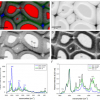
Jan Novotný, Karel Novotný, David Prochazka, Aleš Hrdlička and Jozef Kaiser tell us about “Two dimensional elemental mapping by laser-induced breakdown spectroscopy”. LIBS seems to be finding increasing applications and to be receiving interest by the instrument manufacturers at present. The article provides an introduction to the technique and goes on to show how it can be used for elemental mapping in materials analysis.
“Rheo-nuclear magnetic resonance spectroscopy: a versatile toolbox to investigate rheological phenomena in complex fluids” is Claudia Schmidt’s topic. Rheology is an important science, and NMR has a number of uses within it. However, challenges remain for the simultaneous measurement of rheological and NMR parameters.
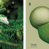
“Elucidating structural and compositional changes in plant tissues and single cells by Raman spectroscopic imaging” is the topic of the next article by Batirtze Prats Mateu, Barbara Stefke, Marie-Theres Hauser and Notburga Gierlinger. Understanding plant cells is important for the best use of plants in traditional and new applications. Raman spectroscopic imaging represents one of the best ways to unravel the molecular structure in the native environment of plant tissues.
Hans Lohninger and Johannes Ofner describe “Multisensor hyperspectral imaging as a versatile tool for image-based chemical structure determination”. They describe the features of a software package that allows the combined analysis of hyperspectral data from different imaging techniques. This multisensor approach providing complementary information has many advantages.
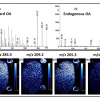
Robert Bradshaw and Simona Francese tell us about “Matrix–assisted laser desorption ionisation tandem mass spectrometry imaging of small molecules from latent fingermarks“. Especially when looking at small molecules in fingermarks, isobaric species can be a problem and this has the potential to affect the outcome of any court case if not handled appropriately. Tandem mass spectrometry can be used as an alternative to high-resolution MS and ion mobility.
“Shedding light on plant biology by Fourier transform infrared spectroscopy of pollen” by Boris Zimmermann and Achim Kohler. Currently, pollen identification is mostly done under a light microscope. FT-IR spectroscopy of pollen grains provides rapid and simple identification of pollen, with the added benefit of providing environmental information.
Orthogonal spectroscopic techniques for the early developability assessment of therapeutic protein candidates” are described by Patrick Garidel, Anne Karow and Michaela Blech. Due to its cost and time implications, in the early development phase of drug discovery the use of othogonal techniques, based on different physical observables, is important for correct decision-making.
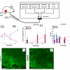
Mid-infrared spectroscopic imaging is a rapidly emerging technique in biomedical research and clinical diagnostics that takes advantage of the unique molecular fingerprint of cells, tissue and biofluids to provide a rich biochemical image without the need for staining. Spectroscopic analysis allows for the objective classification of biological material at a molecular level.1 This “label free” molecular imaging technique has been applied to histology, cytology, surgical pathology, microbiology and stem cell research, and can be used to detect subtle changes to the genome, proteome and metabolome.2–4
Another area of application of XRF, “Determination of elemental distribution or heterogeneity by X-ray fluorescence”, is considered by Christopher Shaffer and Didier Bonvin. The ability of modern X-ray spectrometers to perform small spot analysis as well as mapping has opened up new applications in non-homogeneous samples. The authors show applications in metals, precious alloys as well as rocks.
Knowledge about the particles in the air is important because of their effect on our health and their impact on our climate through cloud formation and transport of nutrients into the oceans. Ursula Fittschen describes “Strategies for ambient aerosols characterisation using synchrotron X-ray fluorescence: a review”. This technique can provide elemental determination and speciation of aerosol particulates with limits of detection in the pg m–3 range for many elements.
The analysis of turbid samples is increasingly important, not least due to their widespread occurrence in natural samples. Dmitry Khoptyar, Sören Johansson, Staffan Strömblad and Stefan Andersson-Engels show “Broadband photon time-of-flight spectroscopy as a prospective tool in biomedicine and industrial process and quality control”. The authors describe their recent development of a broadband spectrometer for evaluation of absorption and scattering spectra of very diverse turbid materials in the visible and close-near infrared (NIR) regions and its application with milk, cheese and paper samples.

It is important that places of archaeological and architectural importance need to be explored without damage. Atomic Dielectric Resonance (ADR) can be used for identifying sub-surface geological features. This technology1 uses a novel coherent beam, which has been used in the oil, gas and water industries, to provide information on what lies beneath the earth’s surface, without the need to drill cores.

“Spectral database for postage stamps by means of FT-IR spectroscopy” by Eleonora Imperio, Gabriele Giancane and Ludovico Valli will be of great interest. As well as helping to detect forgeries, FT-IR has been used to create a database which also charts the history of the technology used to create stamps. Quite rightly, they are considered by many to be works of art.
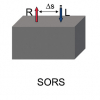
“Emerging sampling approaches for Raman analysis of foods” by Nils Kristian Afseth, Matthew Bloomfield, Jens Petter Wold and Pavel Matousek describes how a number of instrumental developments are enabling Raman spectroscopy to find increasing applications in food analysis. They show how Spatially Offset Raman Spectroscopy (SORS) is being used to analyse quality parameters in salmon, including the content of fat, its fat composition and the content of carotenoids. Traditionally, the preserve of NIR spectroscopy, Raman may increasingly be used for the analysis of food and other biological matrices.
“Membrane inlet mass spectrometry for in situ environmental monitoring” by Simon Maher, Fred Jjunju, Iain Young, Boris Brkic and Stephen Taylor looks at a technique that is 50 years old but is now being applied for field analysis. As well as a brief overview of the technique, they show how it can be used to monitor oil-in-water levels before discharge from oil termini.
Malvina G. Orkoulaa and Christos G. Kontoyannisa,b
aDepartment of Pharmacy, University of Patras, Rio-Patras, Greece. E-mail: [email protected]
bICE-HT/FORTH, PO Box 1414, University Campus, Rio-Patras, Greece
The authors give us a “Review of nanoscale infrared spectroscopy applications to energy related materials”. Fuel cells, photovoltaics and specialised polymers for fracking are all considered.
Mathieu Duval raises the question “Dating fossil teeth by electron paramagnetic resonance: how is that possible?”. Whilst we are all familiar with 14C dating, the use of EPR is less well known. In fact, there are less than 10 laboratories in the world able to carry out EPR dating of fossil teeth!